nikon small world
nikon small world
Small World is regarded as the leading forum for showcasing the beauty and complexity of life
as seen through the light microscope. For over 30 years, Nikon has rewarded the world’s best photomicrographers
who make critically important scientific contributions to life sciences, bio-research and materials science.
http://www.nikonsmallworld.com/
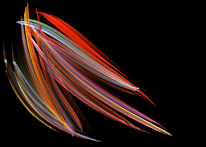
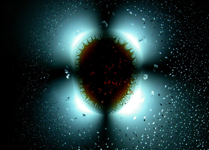
Michael Stringer_Westcliff-on-Sea – Essex, United Kingdom
Specimen: Pleurosigma (marine diatoms) (200x)
Technique: Darkfield and Polarized Light
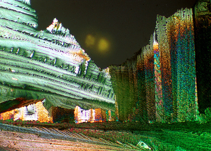
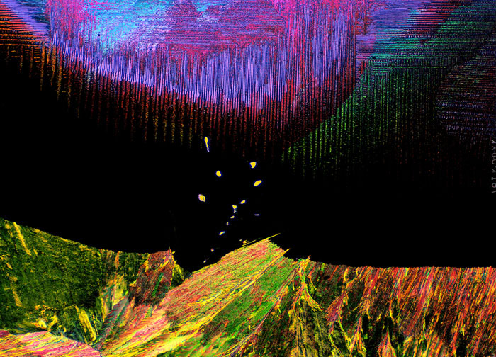
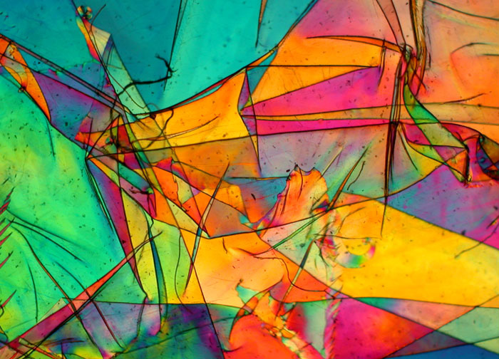
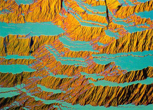
John Hart_Department of Atmospheric and Oceanic Sciences – University of Colorado – Boulder, Colorado, United States
Specimen: Crystallized mixture of resorcinal, methylene blue, and sulphur (13x)
Technique: Polarized transmitted light
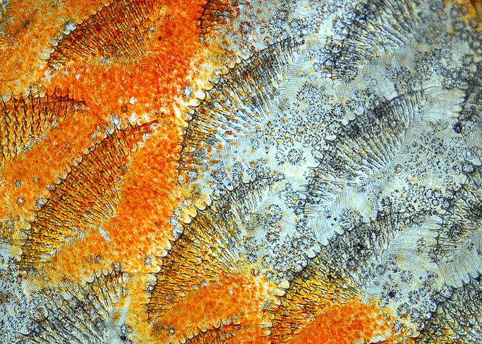
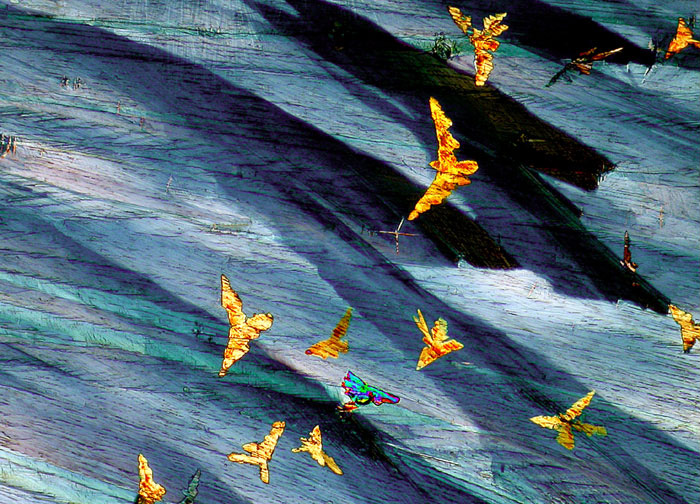
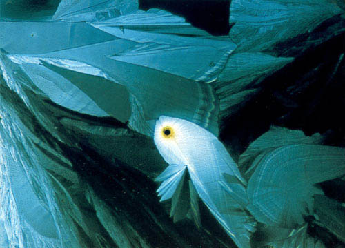
Dr. Havi Sarfaty_Israel Veterinary Association – Ramat-Gan, Israel
Specimen: Scales of Discus fish (20x)
Technique: Transmitted Light
Dr. Margaret Oechsli_Jewish Hospital, Heart & Lung Institute – Louisville, Kentucky, United States
Specimen: Mitomycin (anti-cancer drug) (10x)
Technique: Polarized light
Dr. Rebekah HeltonUniversity of Delaware- Newark, Delaware, United States
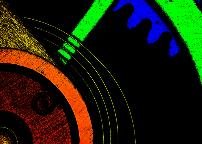
Specimen: Stopwatch (2.5x)
Technique: Confocal
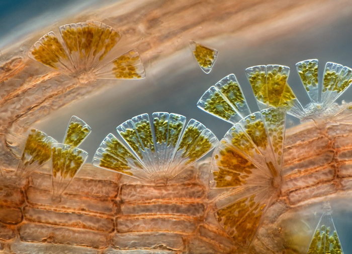
Charles Krebs_Charles Krebs Photography – Issaquah, Washington, USA
Specimen: Marine diatoms attached to Polysiphonia (red algae) (100x)
Technique: Differential Interference Contrast
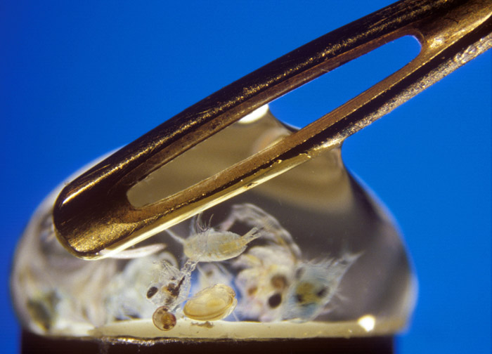
Peter Parks_Imagequestmarine.com – Witney, Oxon, United Kingdom
Specimen: Sea water with mixed zooplankton and needle eye (20x)
Technique: Reflected light
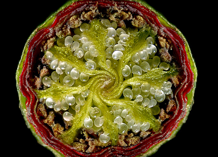
Shamuel Silberman_Ramat Gan, Israel
Specimen: Papaver subpiriforme (corn poppies) flower bud (20x)
Technique: Stereomicroscopy
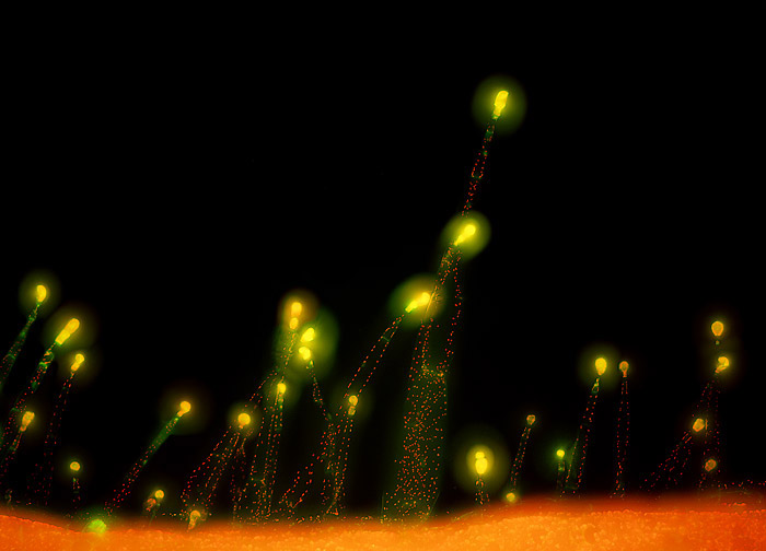
Dr. Heiti PavesTallinn_University of Technology – Tallinn, Estonia
Specimen: Transgenic Nicotiana benthamiana (tobacco) (10x)
Technique: Fluorescence
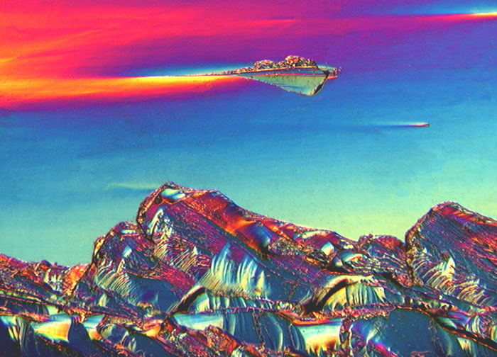
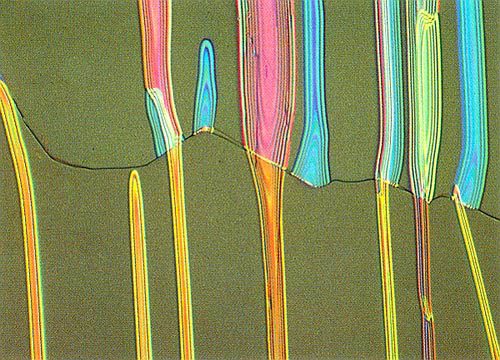
Dr. Oleg D. Lavrentovich_Liquid Crystal Institute – Kent State University – Kent, Ohio, USA
Specimen: Thin nematic film (liquid crystals) (200x)
Technique: Polarized light
Charles B. Krebs_Charles Krebs Photography – Issaquah, Washington, USA
Specimen: Muscoid fly (house fly) (6.25x)
Technique: Reflected light
Stefan Eberhard_Complex Carbohydrate Research Center – University of Georgia – Athens, Georgia, USA
Specimen: Crystallized Vitamin A (40x)
Technique: Polarized light
Edy Kieser_Ennenda, Switzerland
Specimen: Crystallized succinic acid and urea (50x)
Technique: Polarized light
Edy Kieser_Ennenda, Switzerland
Specimen: Crystallized potassium chlorate (40x)
Technique: Polarized light
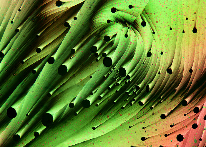
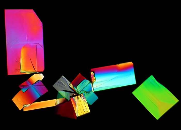
John E. Hart_Program in Atmospheric and Oceanic Sciences, University of Colorado – Boulder, Colorado, USA
Specimen: Crystallized acetaldehyde and carbon tetrabromide (7x)
Technique: Polarized light
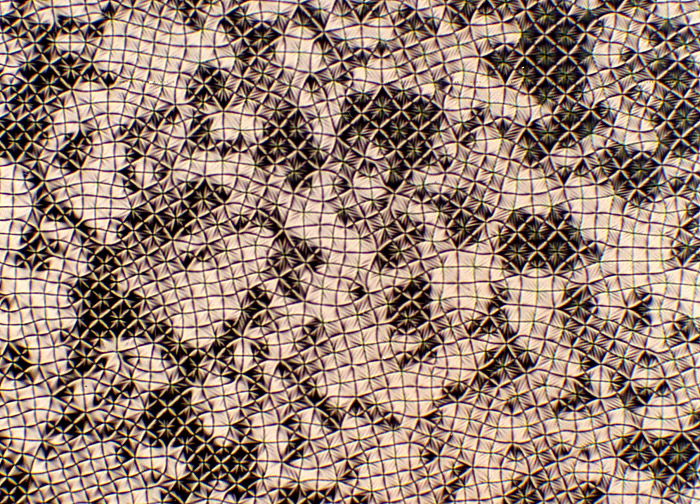
Dr. Christian Bohley_Department of Experimental Physics – Otto-von-Guericke-University of Magdeburg – Magdeburg, Germany
Specimen: Cholesteric phase of 55% CB15 in E48 (substance used in manufacture of Liquid Crystal Displays) (100x)
Technique: Polarized light
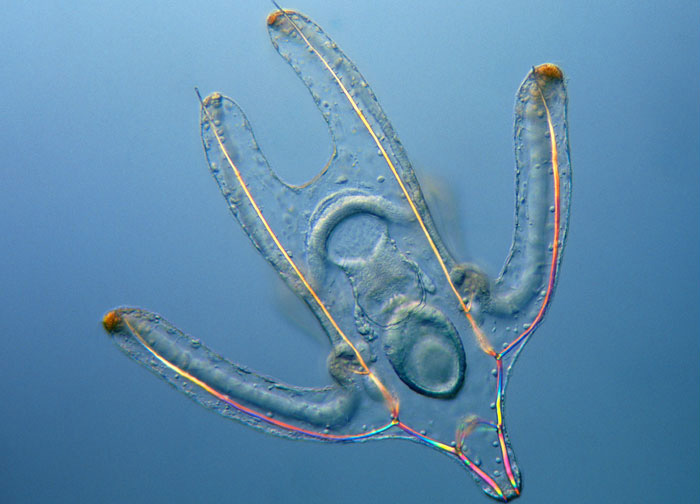
Wim van Egmond_Micropolitan Museum – Rotterdam, The Netherlands
Specimen: Brittle Star Larva, living specimen (100x)
Technique: Differential interference contrast
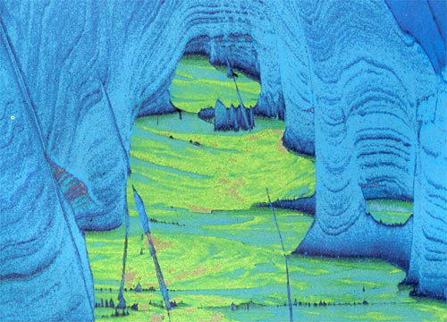
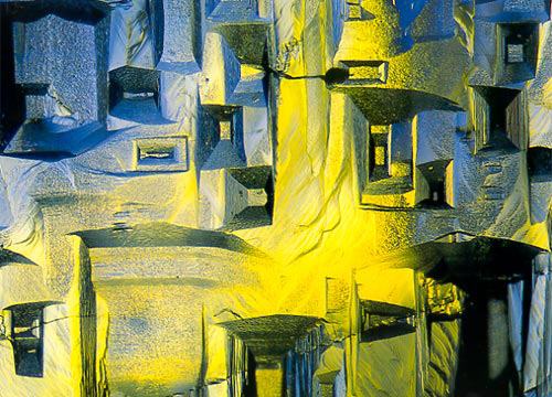
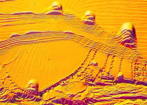
Dr. Lynn A. Boatner and Hu F. Longmire_Oak Ridge National Laboratory – Oak Ridge, Tennessee, USA
Specimen: Surface of titanium carbide crystal (64x)
Technique: Differential interference contrast
Ian C. WalkerHuddersfield, UK
Specimen: Ascorbic acid (Vitamin C) (63x)
Technique: Polarized light
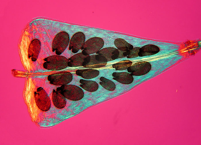
Aaron Messing_The Virginia Company, Inc. – West Orange, New Jersey, USA
Specimen: Embryo seeds of Capsella bursa-pastoris within fruit capsule (25x)
Technique: Polarized Light
Zdenka Jenikova Czech Technical University – Prague, Czech Republic
Specimen: Deformation of a polyethylene folio (40x)
Technique: Polarized Light
Dr. Taijin Lu_Gemological Institute of America – Carlsbad, California, USA
Specimen: Synthetic topaz crystal surface (50x)
Technique: Darkfield
Stefan Eberhard_Complex Carbohydrate Research Center – University of Georgia – Athens, Georgia, USA
Specimen: Acetylsalicylic acid (aspirin) melted with sulfur (40x)
Technique: Polarized Light
Anna S. Teetsov_McCrone Associates, Inc. – Westmont, Illinois, USA
Specimen: Polypropylene (a plastic) melted with phthalocyanine blue pigment (50x)
Technique: Polarized Light
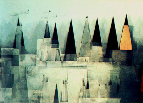
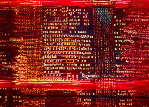
Ulrich Büttner_Daimler Benz Research Center – Ulm, Germany
Specimen: Indium phosphide surface (1000x)
Technique: Brightfield / DIC
Lars Bech_Naarden, The Netherlands
Specimen: Doxorubin in methanol and dimethylbenzenesulfonic acid (80x)
Technique: Polarized Light
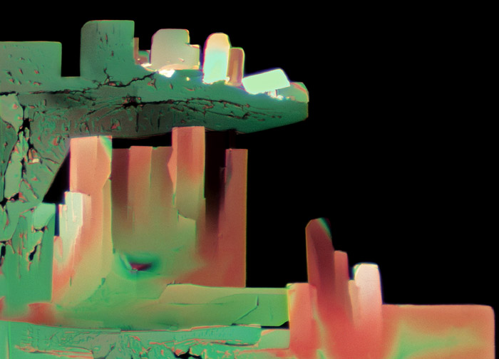
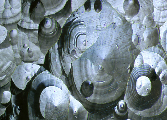
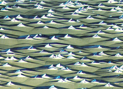
John I. Koivula_Gemological Institute of America – Santa Monica, California, USA
Specimen: Growth steps on the face of a quartz crystal (40x)
Technique: Differential Interference Contrast
Derek Hull_Department of Materials Science and Engineering – University of Liverpool -Liverpool, England
Specimen: Fracture surface of mica (100x)
Technique: Brightfield
Dr. Kari A. KinnunenGeological Survey of Finland – Espoo, Finland
Specimen: Gem beryl crystal (8x)
Technique: Rheinberg Illumination
Richard H. LeeArgonne National Laboratory – Argonne, Illinois, USA
Specimen: Crystals evaporated from solution of magnesium sulfate and tartaric acid (50x)
Technique: Polarized Light
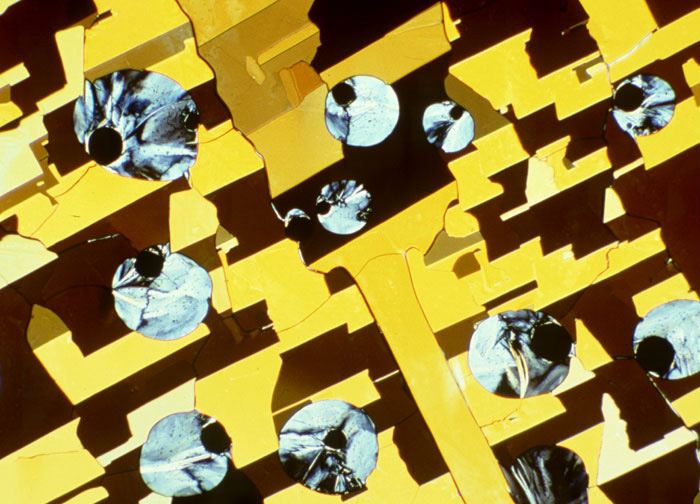
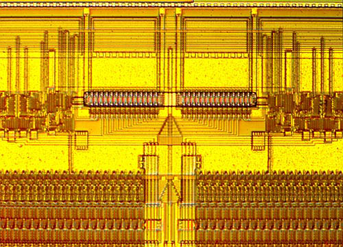
Karl E. Deckart_Eschenau, West Germany
Specimen: Structure of 1 megabite integrated circuit (40x)
Technique: Differential Interference Contrast
James BellMelrose, Massachusetts, USA
Specimen: Stained section of Sequoia redwood (125x)
Technique: Polarized Light
Jean-Claude WittmannCentre National de la Recherche Scientifique – Strasbourg, France
Specimen: Thin crystals of an aromatic hydrocarbon deposited on a glass slide from solution.
Color produced by insertion of mica sheet (165x)
Technique: Polarized Light
David GnizakIn_dependence, Ohio, USA
Specimen: Gold, vaporized in a tungsten boat, in a vacuum evaporator (55x)
Technique: Vertical Illumination – Normarski Differential Interference

